The Neck Injury Your Doctor Might Be Missing After an Auto-Accident – Chiropractor in Wellington
The Neck Injury Your Doctor Might Be Missing After an Auto-Accident
New Imaging Methods May Identify Injuries Missed by Conventional Studies
Lots of people understand that whiplash, neck pain, headaches, and pinched nerves can occur following a car crash. In fact, that’s one of the most common reasons that people begin traditional chiropractic care.
Most people will recover from an accident without any problems. However, some patients may begin to develop chronic symptoms of whiplash, and others may start showing worsening, unexplained symptoms months or years later. There are some new imaging techniques that may help explain why.
Upright MRI vs Recumbant MRI: Gravity exposes weaknesses
If you’ve ever had an MRI done, chances are you’ve had this imaging done while lying down. Most MRI’s have you lying on your back for about 30 minutes while it hums away taking images. This allows you to minimize how much you move which will distort the images.
In more recent years, MRI machines have been developed that allow you to see the spine in a seated or standing position. Why is this important?
- Most people with spine problems don’t complain about their pain while lying down. Their pain is triggered when they are standing or sitting in gravity. People need to be evaluated in the situation that causes their dysfunction.
- Gravity can force the soft tissues of the body to deform and show what’s really going on. Placing the body under stress allows you to see what’s happening that can be triggering pain or dysfunction.
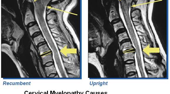
Some more recent studies have shown that various spinal conditions can show up during an Upright MRI that are frequently missed on a supine (laying down) MRI. This can include increased disc herniation, spondylolisthesis, and lowered brain tissue in scoliosis patients (tonsilar ectopia). Check out the 2 examples below:
Neck

A borderline normal MRI suddenly has some noticeable changes in the brain and disc just by getting a person from lying down to sitting up. Image Credit to FONAR
Lower Back
This is one of the major reasons why I don’t recommend invesion or traction therapy on a regular basis. Traction and inversion helps people out of gravity, but what happens when they are upright again?
We live our lives in a gravitational field. In order to truly help someone function better, we must take care of them in a way that promotes optimal function when they are upright.
The primary criticism of upright imaging is that the resolution of the image isn’t as high as standard imaging.
What Does This Mean For You As A Patient?
“One of the most frustrating things you can hear from a doctor is that nothing is wrong”
Lots of patients go to the doctor with various problems after an accident. Here are some of the more common post-accident problems I see in my Wellington office:
- Arm pain
- Headache
- Dizziness
- Brain fog
- Whole Body Pain
- Jaw pain/TMJD
For many of these patients, diagnostic testing and imaging comes out negative, and it leaves the doctor stumped as to what may be causing their problem. A negative test can be devastating for a chronic pain or chronically ill patient. At the very least, you want the peace of mind of knowing that there’s a CAUSE for your pain and suffering. An explanation and closure can be extremely therapeutic for many.
There are 4 main neck injuries that can be spotted on upright MRI that can be missed on standard MRI:
- Moderate to Severe Disc Herniation/Pinched Nerves – neck pain, arm pain, shoulder blade pain
A lot of patients have symptoms of a pinched nerve in their neck, but standard MRI shows that the nerves are okay. When you put the spine in an upright position, gravity can put the additional pressure onto the intervertebral discs needed to push the disc into the nerve roots causing your symptoms.This is especially important considering that most pinched nerve symptoms happen when you are upright, not when you’re lying down. - Arnold-Chiari Malformation/Cerebellar tonsilar ectopia – headaches, dizziness, visual disturbances
Cerebellar tonsilar ectopia is a condition in which a small piece of your brain starts to get pushed down towards the neck. I know it sounds like something crazy that should probably be fatal, but the truth is that thousands of people have this condition and don’t show any obvious signs or symptoms.However, some patients with this problem will show symptoms like headache, drop attacks, dizziness, and balance problems. This is what it looks like on MRI:
Left shows normal brain placement. The right is the same patient in a seated positio showing the brain being pushed down towards the neck.
A study published in 2013 showed that more people may have this problem than originally thought. Upright MRI showed that the incidence of this problem increases to almost 25% of patients who have had whiplash have this issue, where only 5% of people have it with no injury history.
The problem is that this piece of brain tissue can block fluid from moving normally in the brain. When this fluid blockage happens, the brain starts to experience increased pressure, and nutrients fail to circulate properly.
- Damage to Upper Cervical Ligaments – neck pain, headaches, painful Range of motion, chronic pain
If you have ever had a sprained ankle, then you are intimately familiar with the problems associated with ligament damage. Ligaments are the glue that hold your joints together. But they are also tissues that transmit a lot of information to the brain and nervous system. When you have ligament damage it makes joints less stable, induces swelling, and makes the joint more sensitive to pain.Most radiologists will look at big bulky ligaments for signs of damage. Doctors of the upper cervical spine will look for damage to the small ligaments that connect your neck to your head. These include the alar, transverse, atlanto-occipital membrane, and atlanto-axial membrane.These are the ligaments that can create a problematic shifting of the spine that Structural Chiropractors see on a regular basis. Damage to these ligaments also make the spine less stable, and usually require more frequent visits to get a more stable response post-injury. - Rotational Atlas Displacement with venous obstruction – postural distortion, decreased concentration, chronic neural degeneration
This is the area of expertise for chiropractors that focus on the top vertebra in the spine called the Atlas.When there is sufficient damage done to the ligaments of the neck from the force of an accident or collision, this ring of bone at the top of the neck can shift out of normal ranges. This additional shifting of the atlas can encroach the vessels that drain the brain of blood and spinal fluid.
Although the theory is still controversial, blockage of venous blood leaving the brain has been tied to MS, Meniere’s Disease, Parkinson’s Disease, and migraine headache.
Are These Neck Problems Correctable?
As of now there are only case studies and anecdotes utilizing pre and post correction upright imaging, so it’s difficult to say. However, many of these pre and post images are compelling showing things like:
- Reduction/resolution of herniated/protruded discs
- Reduction in tonsilar ectopia
- Reduction in Atlas Displacement
I’ve been learning more about these images, and there are even a couple of imaging centers here in Palm Beach County that take these upright views. Only a few chiropractors are making use of these imaging techniques to deliver care. There’s a lot of work that needs to be done before this type of imaging can be used by more chiropractors, but some of the initial reports have been encouraging.
The good news, chiropractors that focus on Atlas Displacement Complex are accomplishing some of the same clinical outcomes utilizing precision upper cervical x-ray set ups. While this type of imaging may help us improve the quality of care for difficult cases, it also confirms that upper cervical doctors have been on the right path all along.


Leave a Reply
Want to join the discussion?Feel free to contribute!